loader 1
Reimagining breast conserving surgery by operating precisely
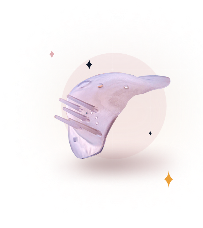
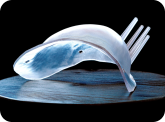
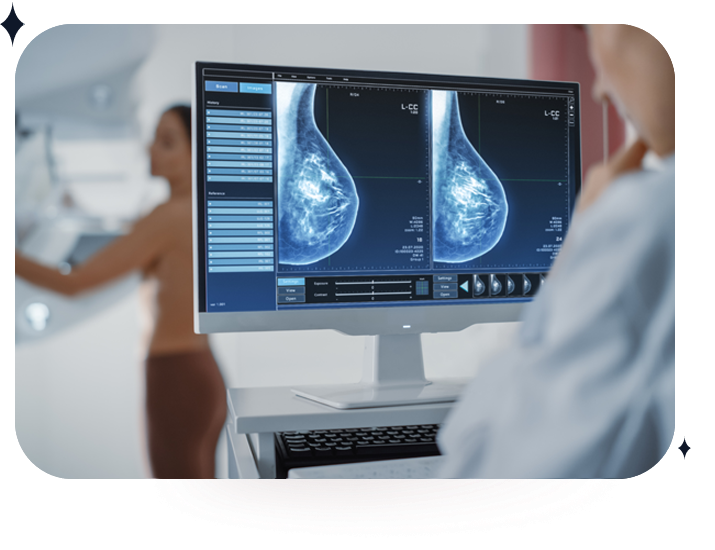
The Breast Cancer Locator™ System (BCL™) is a proprietary, patient-specific, 3D printed surgical guide designed to reduce positive margins during breast cancer tumor removal. The BCL™ and Visualizer™ provides information regarding tumor size, shape, and margin boundary to assist surgeons in the excision of cancer and preserve normal breast tissue.
CairnSurgical receives patient MRI data, analyzes it to define tumor geometry and location, and uses the data to fabricate the BCL™.
 Try Visualizer™ App
Use the right mouse button to rotate and the mouse scroll wheel to zoom. Experience is optimized for Chrome and Firefox browsers.
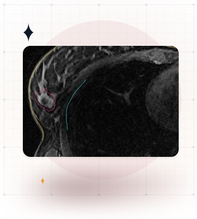
After the surgeon and patient choose a lumpectomy plan, the patient receives a supine (patient facing up) MRI in the same position as during the surgery, which makes the image more accurate than face down.
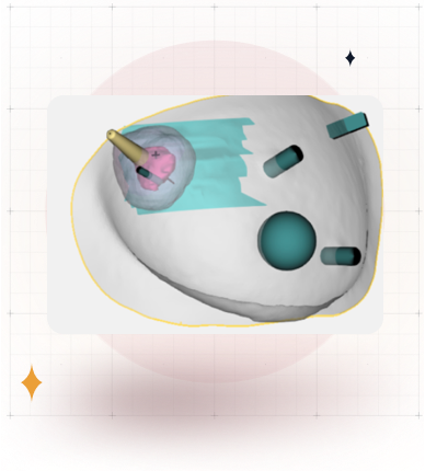
We develop a patient-specific planning guide and design based on the supine MRI. The Visualizer™ 3D interactive visualization is generated for the patient. The Visualizer ™ is a 3D image of the tumor and surrounding tissue inside the breast. The visualization link is sent to the surgeon.
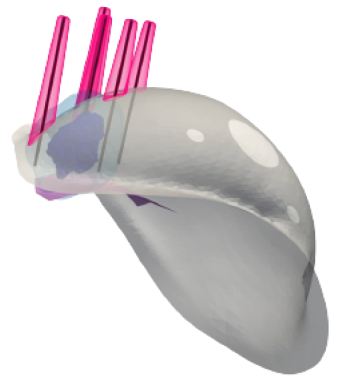
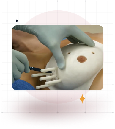
The MRI is used to print the BCL™ - a polymer guide matched to the dimension of the patient’s breast with guiding ports to bracket the tumor. The BCL™ is shipped to the surgeon.
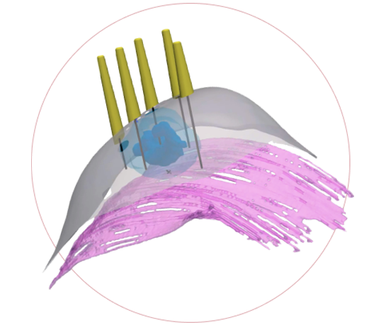
After the patient is under general anesthesia, the surgeon places the BCL™, marks the projected edges of the cancer on the skin surface, and places four bracketing wires 1 cm from the MRI-defined tumor volume. The BCL is removed, and surgery is performed with guidance from the wires and the Visualizer™.
The BCL™ precisely defines the extent of the tumor and excision volume, not just a center point or edge point. The Visualizer™ provides a 3D image of the tumor in the supine position including volumetric guidance with in-vivo cues.
Intuitive, not complicated, no added capital equipment.
No need for pre-surgery wire or seed insertion. BCL™ is used during surgery after general anesthesia has begun.
Risk of re-excision surgery significantly reduced.
The BCL™ precisely defines the extent of the tumor and excision volume, not just a center point or edge point.
The Visualizer™ provides a 3-D image of the tumor in the supine position including volumetric guidance with in-vivo cues.
Intuitive, not complicated, no added capital equipment.
No need for pre-surgery wire or seed insertion. BCL™ is used during surgery after general anesthesia has begun.
Risk of re-excision surgery significantly reduced.


Professor of Surgery, Dartmouth Geisel School of Medicine
In a recently completed Breast Cancer Locator (BCL) clinical study in Europe at five hospitals with nine surgeons, only two out of 33 patients treated per protocol had positive margins, or 6%.
A randomized, prospective clinical trial of the BCL™ is underway, offering hope for surgeons and breast cancer patients
alike. The goal of the clinical trial is to improve the accuracy of tumor removal, resulting in more negative margins and
fewer re-excision surgeries.
CairnSurgical Inc. 16 Cavendish Court, Lebanon NH 03766