loader 1

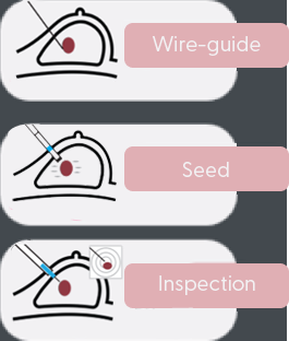
All probes based on a point in space
One point in space for guidance to tumor center
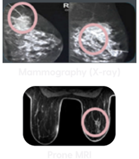
Images don't match breast geometry in OR

First volume-based 3D interactive guidance tool
3D guidance to detect location, size, and shape of tumor – superior to single point guidance

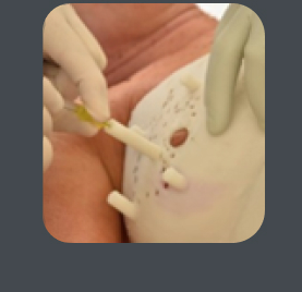
Wires in center and surrounding the tumor
To identify the tumor plus clear margin – superior to a single wire or seed
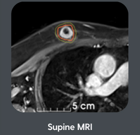
Images taken in surgical position
Images that inform for the surgical position – unlike mammograms and prone (patient lying face down)
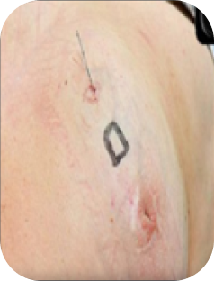
Addressing the needs of every patient and unique tumor shapes. Our breast cancer locator is molded to the actual shape of a patient’s breast which enables the surgeon to place the markers and accurately bracket the tumor.
Markers are placed while patients are under anesthesia. Patients don’t experience the typical pain associated with traditional wire localization procedures.
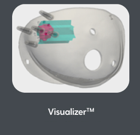
The CairnSurgical BCL™ system consists of the BCL™ and the Visualizer™. With our system, there are no additional capital expenditures for medical equipment and no need for a different procedure where the radiologist places a wire or a seed.
The Visualizer™ guides the surgeon to the precise tumor boundaries and margins, helping establish an accurate surgical plan.
From the supine MRI data, Cairn Surgical creates The Visualizer™ - an interactive a web-based program that depicts a 3D model of the tumor, which can be adjusted by the surgeon to show the shape of the tumor through various specific views identifying dimensions, tumor location, and a darker boundary around the tumor that represents a 1 cm margin. The Visualizer™ also features measurements of the distance from the chest wall to the tumor and from the skin surface to the tumor. The Visualizer™ can be used for pre-planning ahead of surgery and serves as a reference during the surgery.
A lumpectomy is entirely successful when a surgeon achieves clean margins around the excised tissue. Clean in this context means that the tissue on the surface of the specimen contains no cancer cells. This is challenging to accomplish since surgeons are rarely guided by the kind of intraoperative imaging (MRI) that would allow them to clearly see the tumor. To increase the odds of success, some surgeons sometimes shave off an additional millimeter around the tumor for good measure, but that doesn’t guarantee clean margins. The BCL™ and the Visualizer™ provide surgeons a more accurate view of the tumor before, during and after surgery greatly increasing the chance of delivering clean margins and thus, better post-operative breast preservation & cosmetic outcomes.
Chief of Surgery, Women & Infants Hospital, RI
Professor of Surgery, Brown University
Chair, Department of Breast Oncology Moffitt Cancer Center
St. Peter’s Health Partners Albany, NY

A Multi‑institutional Study to Evaluate the Effectiveness and Safety of a Supine MRI‑Based Guidance System, the Breast Cancer Locator™, for Breast Conserving Surgery in Patients with Nonpalpable Breast Cancer. Annals of Surgical Oncology 2025
A total of 35 subjects were enrolled at 5 sites by 9 surgeons. In the 33 patients treated per protocol, 31 had margin negative resections (94%). All 31 patients with negative margins had negative margins on the primary lumpectomy specimen resected with BCL guidance. Additional shave margins were taken in 4 of the 31 patients; no cancer was present in the shaves. A total of 25 patients had invasive ductal carcinoma, 7 invasive lobular carcinoma, and 3 ductal carcinoma en situ (DCIS).
A pilot multi-institutional study to evaluate the accuracy of a supine MRI based guidance system, the Breast Cancer Locator™, in patients with palpable breast cancer. Surgical Oncology 2022
Fourteen patients were enrolled at 4 different sites by 6 surgeons. BCL™'s were successfully manufactured for all patients. The central wire was deployed within the tumor on specimen mammogram in 12 of the 13 patients who had a central wire placed (92%).
The cancer was excised with negative margins in 14/14 cases (100%). No adverse events occurred.
A Patient-Specific 3D-Printed Form Accurately Transfers Supine MRI-Derived Tumor Localization Information to Guide Breast-Conserving Surgery. Annals of Surgical Oncology Oct 2017
Clear tumor margins were achieved in all 19 patients. Resection volumes were comparable to optimal resection volumes determined by tumor modeling based on the supine MRI.
“The shape of breast cancer” Springer Nature, July 2020
Information obtained from a supine MRI can be used to generate 3D tumor models and rapidly classify breast tumor
shapes. The vast majority of invasive cancers and DCIS are not spherical. Knowledge of tumor shape may allow
surgeons to excise breast cancer more precisely.
CairnSurgical Inc. 16 Cavendish Court, Lebanon NH 03766