loader 1
of women develop invasive breast cancer during their lifetime
of breast cancer tumors are irregularly shaped making it difficult for surgeons to accurately locate the whole tumor in order to remove it.
of women with breast cancer must undergo a second surgery because some of the cancer is missed

An MRI with the woman lying on her back is done with the breast positioned as it will be during surgery, so the exact detail of the size, shape, depth, and edges of the tumor can be seen.
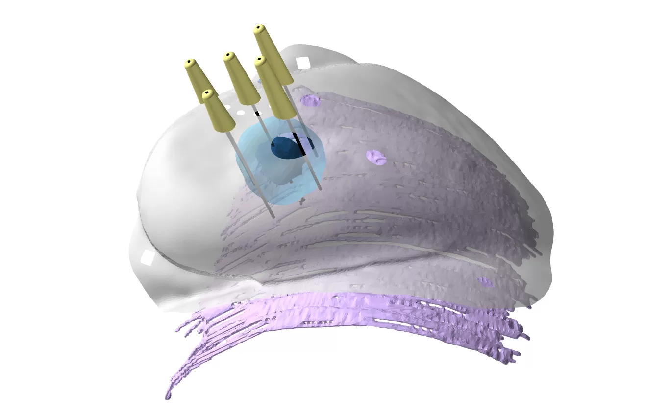
This image is used to analyze the tumor and build a three-dimensional model of the tumor and breast. This offers the surgeon the view of the breast and tumor in the same position as it will be during surgery.

A 3D-printed bra-like form (BCL™) that fits the unique shape of the patient’s breast is made. It has markers for locating the tumor and is placed on the patient while she is asleep during surgery. The BCL™ design guides the surgeon to the precise tumor edges.

An interactive 3D image – the Visualizer™ offers visualization of the tumor and its surroundings, allowing the surgeon to
view the tumor from all angles, including the size, shape, and distance of the tumor from the skin and chest wall.
This enables the surgeon to see the best position for surgery. It is the only tool available that shows these views for
breast conserving surgery.
Patient, Clinical Trial